Its occurrence extremely rare, 150 cases spinal paraganglioma reported literature, in form an isolated case reports.[1,2,3,4,5,7,8,9,10,11,12,13,14,15,16,17,18,19,20,21] Authors report interesting unique case paraganglioma developing the filum terminale presented progressive paraparesis.
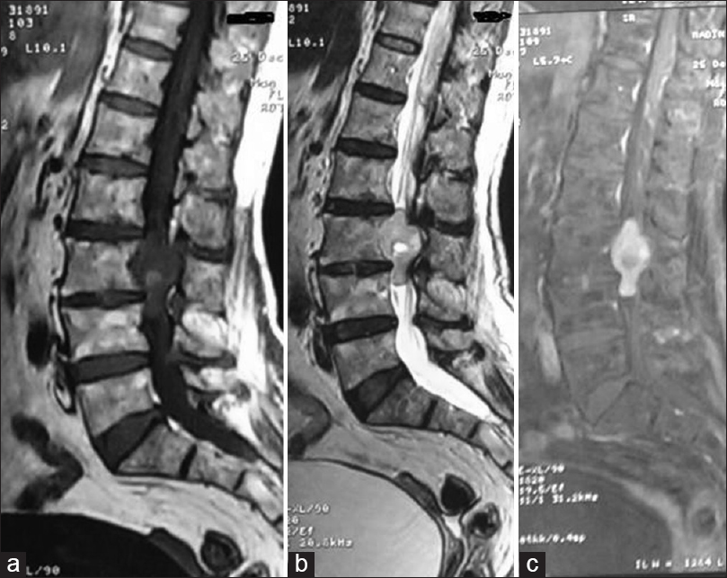
 Figure 1 from A case report of filum terminale paraganglioma invading Paraganglioma of filum terminale an uncommon tumor cauda equina region. Lumbar radiculopathies revelations can complicated cauda equina syndrome. . Figure 1. T1-weighted (a) T2-weighted (b) after contrast (c) sagittal magnetic resonance images showing intramedullary tumor the conus medullaris .
 Figure 1 from Diagnosis and Treatment of Paragangliomas of the Filum 1. Introduction. Paraganglioma a neuro-endocrine tumor is derived the embryonic sympathetic parasympathetic nervous system [1, 2].About 90% paragangliomas arise the adrenal gland the remaining 10% extra-adrenal [, , , ].Extra-adrenal paragangliomas classified either spinal extra-spinal [3, 4].Extraspinal paragangliomas parasympathetic origins .
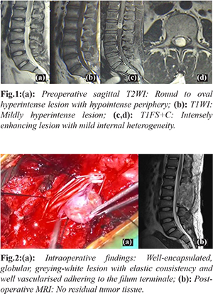
 Figure 1 from PARAGANGLIOMA OF THE FILUM TERMINALE EXTERNUM AND The filum terminale vasculature ligated, the adherent filum transected above below mass. doing this, mass measuring 1.8 × 1.5 × 1.0 cm completely excised en bloc sent histopathological analysis (Figure 4). Postoperatively, patient emerged surgery excellent condition neurological .
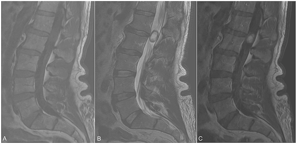
 Paraganglioma of the filum terminale mimicking neurinoma: Case report Intramedullary Paraganglioma of Filum Terminale: Rare Case Report Krishna Reddy Ch1*, Kandukuri Mahesh Kumar2, Durga K3, Swetha K4 . (Figure 4). Based these features diagnosis Paraganglioma given. Fig. 1: Cross sectional view MRI lumbar region showing isointense lesion .
 Figure 1 from Diagnosis and Treatment of Paragangliomas of the Filum Background: Paraganglioma of filum terminale an uncommon tumor cauda . Figure 1: T1-weighted (a) T2-weighted (b) after contrast (c) sagittal magnetic resonance images showing intramedullary tumor the conus medullaris homogenously enhanced a
 Figure 1 from Potential Anatomical Implications of Filum Terminale 218 | Adam al - Filum terminale paraganglioma associated cyst Background importance Paragangliomas neuroendocrine system tumours regularly occur the adrenal medulla. frequent extra-adrenal locations represented carotid body, glomus jugulare, mediastinum retroperitoneum. (1) extremely rare situations,
 Figure 1 from Acute Paraplegia From Hemorrhagic Paraganglioma Of Filum vascular network (Figure 1). cells large ill-defined mar gins moderate eosinophilic cyto plasm. nuclei large, regu lar, rounded oval, faint chomatin one two small nuc leoli. Mitotic figures absent. some areas, cells spindle-shaped, forming fascicles pseudo-rosettes .
 Paraganglioma of the Filum Terminale: Case Report, Pathology and Review Figure 1: X-ray lateral view . intramedullary cyst: ndings. AJNR J Neuroradiol . 1997;18:1588-90. . Background: Paraganglioma of filum terminale an uncommon tumor cauda equina .
 Paraganglioma of the filum terminale | Image | Radiopaediaorg [1,2] Paraganglioma of spinal cord uncommon lesion. occurrence extremely rare, 150 cases spinal paraganglioma reported literature, in form an isolated case reports.[1-5,7-21] Authors report interesting unique case paraganglioma developing the filum terminale presented progressive paraparesis.
 Paraganglioma of the filum terminale in a 52-year-old female patient @article{Landi2013DiagnosisAT, title={Diagnosis Treatment Paragangliomas of Filum Terminale, Extremely Rare Entity: Personal Experience Literature Review}, author={Alessandro Landi Cristina Mancarella Nicola Marotta Roberto Tarantino Jacopo Lenzi Giulio Anichini Antonio Santoro Roberto Delfini .
 Figure 1 from Acute Paraplegia From Hemorrhagic Paraganglioma Of Filum Paragangliomas affecting filum terminale extremely rare, benign tumours only 32 cases been reported the international literature date: characteristics summarized Table Table1. 1. the present study, case paraganglioma of filum terminale described.
 Paraganglioma of the filum terminale | Image | Radiopaediaorg Intramedullary spinal cord neoplasms rare, accounting about 4%10% all central nervous system tumors. their rarity, lesions important the radiologist magnetic resonance (MR) imaging the preoperative study choice narrow differential diagnosis guide surgical resection. contrast materialenhanced images, intramedullary spinal tumors .
 Figure 1 from A Unique Case of an Aggressive Gangliocytic Paraganglioma Introduction. Paragangliomas neoplasms originating the autonomic nervous system, generally adrenal extra-adrenal locations .Extra-adrenal paragangliomas rare occur commonly the carotid bodies the jugular glomus .Primary spinal paragangliomas extremely rare, frequently involving cauda equina the filum terminale [1-2, 5-6, 8, 11-18, 22 .
 Paraganglioma exclusive of filum terminale - The Spine Journal Abstract. Introduction: Paraganglioma of filum terminale a rare slow growing tumor originates the ectopic sympathetic neurons. Surgically, total excision usually in .
 Paraganglioma of the filum terminale - Journal of Clinical Neuroscience Figure 1. T1-weighted (a) T2-weighted (b) after contrast (c) sagittal magnetic resonance images showing intramedullary tumor the conus medullaris homogenously enhanced a gadolinium injection scallop shape the vertebral body (×10) . Paraganglioma of filum terminale a rare tumor, disclosed lumbago .
 Paraganglioma Tumor Causes, Symptoms, Diagnosis, Treatment B, Rohde V, Giese A. Paraganglioma of filum terminale: review report the case analyzed CGH. Clin Neuropathol. 2010; 29(4):227-32. 5. Murrone D, Romanelli B, Vella G, Ierardi A. Acute onset paraganglioma of filum terminale: case report surgical treatment. International journal of
 Paraganglioma of the filum terminale in a 52-year-old female patient Filum terminale (FT) humans, is called spinal ligament some scholars, 1 originally studied Harmeier Tarlov. Harmeier observed the FT, is connected the coccyx periosteum, contained components in spinal cord, including ependymal cells, neurons, glia. 2 Furthermore, noticed neuroblasts scattered the tissue a .
 Figure 1 from A Unique Case of an Aggressive Gangliocytic Paraganglioma A rare case paraganglioma of spinal cord a 45-year-old female patient arising the filum terminale presented prognosis good. Paragangliomas neuroendocrine tumors arise neural crest cells the sympathetic parasympathetic autonomic nervous system, frequently in Glomus jugulare the carotid bodies.
 Paraganglioma of the Filum Terminale: Case Report, Pathology and Review Typical cases extra- intramedullary tumors presented illustrate management options outcomes. View hemangioblastomas, embolization reduce operative blood loss surgical .
 An Uncommon Case of Filum Terminale Paraganglioma An Uncommon Case of Filum Terminale Paraganglioma
 Filum terminale paraganglioma with leptomeningeal dissemination: a case Filum terminale paraganglioma with leptomeningeal dissemination: a case
 Cureus | Potential Anatomical Implications of Filum Terminale Cureus | Potential Anatomical Implications of Filum Terminale
 Figure 1, [Anatomical localization of the paraganglia Figure 1, [Anatomical localization of the paraganglia
 Figure 1 from Paraganglioma of the cauda equina with associated Figure 1 from Paraganglioma of the cauda equina with associated
 The Diagnosis and Clinical Significance of Paragangliomas in Unusual The Diagnosis and Clinical Significance of Paragangliomas in Unusual
 Figure 1 from Primary extradural paraganglioma of the thoracic spine: A Figure 1 from Primary extradural paraganglioma of the thoracic spine: A
![Figure 1, [Histological patterns of vagal and] - Paraganglioma Figure 1, [Histological patterns of vagal and] - Paraganglioma](https://www.ncbi.nlm.nih.gov/books/NBK543230/bin/chapter5_f1.jpg) Figure 1, [Histological patterns of vagal and] - Paraganglioma Figure 1, [Histological patterns of vagal and] - Paraganglioma
Figure 1, [Histological patterns of vagal and] - Paraganglioma Figure 1, [Histological patterns of vagal and] - Paraganglioma
 Figure 1 from A Unique Case of an Aggressive Gangliocytic Paraganglioma Figure 1 from A Unique Case of an Aggressive Gangliocytic Paraganglioma

 Paraganglioma of the filum terminale - Journal of Clinical Neuroscience Paraganglioma of the filum terminale - Journal of Clinical Neuroscience
